
Grade 10 Biology · Strand 1.0 Cell Biology and Biodiversity · Sub-strand 1.3 Cell Structure and Specialisation
These are free study notes for Grade 10 learners, written to the KICD curriculum design and taken from our full Grade 10 Biology notes. This is the first big topic of Senior School Biology, and everything after it (nutrition, transport, respiration) builds on the organelles described here.
Every living thing, from a yeast cell to a giraffe, is made of cells. The cell is the basic unit of structure and function of life. In this topic we compare the light and electron microscopes, prepare temporary slides and estimate cell size, study the parts of plant and animal cells as seen with the electron microscope, see how special cells are suited to their jobs, and follow how cells are organised into tissues, organs and organ systems.
The cell and the microscope
Cell
The smallest unit of a living thing that can carry out the life processes; the basic unit of structure and function of life.
Most cells are far too small to see with the naked eye, so we study them with microscopes. Cell sizes are measured in micrometres: 1 mm = 1,000 micrometres.
Magnification
How many times larger the image is than the real object.
Resolution (resolving power)
The ability of a microscope to show two points that are very close together as separate points. It decides how much fine detail can be seen.
Magnification is not detail
Enlarging a photograph of a cell on a photocopier makes it bigger but not clearer. Only better resolution shows more detail; that is why the electron microscope reveals organelles the light microscope cannot.
Total magnification of a light microscope
Total magnification = eyepiece magnification × objective magnification.
Eyepiece ×10 and objective ×40: 10 × 40 = ×400.
Eyepiece ×10 and objective ×4: 10 × 4 = ×40.
Light and electron microscopes compared
| Feature | Light microscope | Electron microscope |
|---|---|---|
| Source of "light" | Ordinary light | A beam of electrons |
| Lenses | Glass lenses | Electromagnets |
| Magnification | Up to about ×1,500 | Up to ×500,000 or more |
| Resolution | Low (about 0.2 micrometres) | Very high (shows organelles in detail) |
| Specimens | Living or dead, thin and often stained | Dead only, in a vacuum, very thin sections |
| Image | In natural colour | Black and white (colour may be added by computer) |
| Size and cost | Small, cheap, easy to use and carry | Very large, expensive, needs trained staff |
A transmission electron microscope passes electrons through very thin slices and shows the inside of cells. A scanning electron microscope bounces electrons off the surface and gives a three-dimensional view of the surface.
Preparing slides and estimating cell size
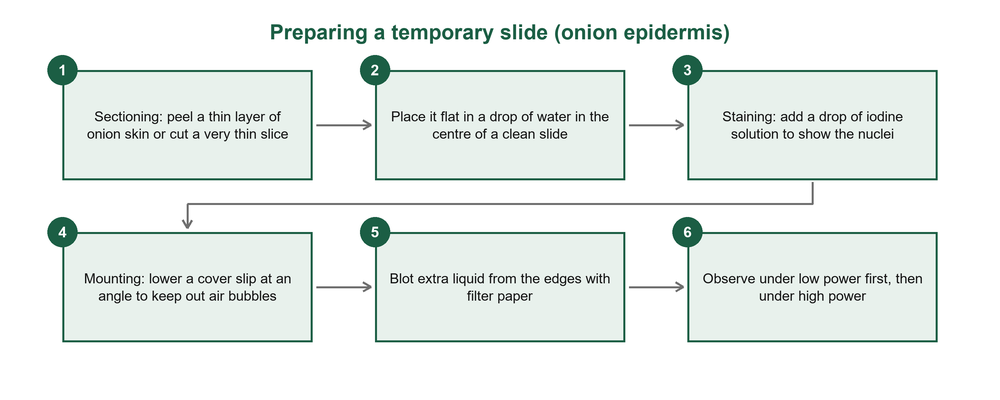
Sectioning
Cutting a specimen into very thin slices so that light can pass through it.
Staining
Adding a dye (such as iodine) to make parts of the cell, especially the nucleus, show clearly.
Mounting
Placing the specimen in a drop of liquid on a slide and covering it with a cover slip.
Fixation
Killing and preserving tissue quickly (usually with chemicals) so its structure does not change; used for permanent slides.
Safety and good practice
Use plant material such as onion bulbs, kale leaves or young stems; do not use human tissue. Lower the cover slip slowly at an angle so no air bubbles are trapped. Always focus first on low power.
Estimating cell size
Using the field of view
- Place a transparent ruler on the stage and focus on low power.
- Measure the diameter of the circle of light (the field of view), for example 2 mm = 2,000 micrometres.
- Replace the ruler with your slide and count how many cells fit across the field of view.
- Divide: size of one cell = diameter of field ÷ number of cells.
Onion cells
Field of view = 2 mm = 2,000 micrometres; 8 cells fit across it.
Size of one cell = 2,000 ÷ 8 = 250 micrometres (0.25 mm).
At ×40 objective (10 times the ×4 power), the field is 2,000 ÷ 10 = 200 micrometres wide.
Magnification of a drawing
Magnification = length of drawing ÷ actual length (same units).
A cell 0.2 mm long is drawn 60 mm long: 60 ÷ 0.2 = ×300.
Actual length = length of drawing ÷ magnification.
The animal cell under the electron microscope
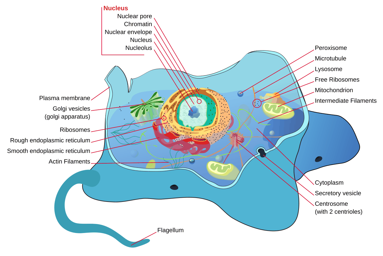
| Organelle | Structure | Function |
|---|---|---|
| Cell membrane | Thin, flexible boundary | Controls what enters and leaves (selectively permeable) |
| Cytoplasm | Jelly-like fluid | Holds organelles; many chemical reactions take place here |
| Nucleus | Double membrane with pores; chromatin and a nucleolus | Controls the cell; carries genetic material (DNA) |
| Mitochondrion | Rod-shaped; inner membrane folded into cristae | Site of aerobic respiration: releases energy |
| Ribosomes | Tiny granules, free or on rough ER | Make proteins |
| Endoplasmic reticulum | Network of membrane sacs; rough (with ribosomes) or smooth | Transports materials; smooth ER makes lipids |
| Golgi apparatus | Stack of flattened sacs with vesicles | Modifies, packages and releases proteins (secretion) |
| Lysosomes | Small sacs of digestive enzymes | Break down worn-out organelles and engulfed germs |
| Centrioles | Two short tubes near the nucleus | Help in cell division |
Link structure to activity
Muscle and sperm cells have many mitochondria because they need a lot of energy. Gland cells that make enzymes or hormones have a lot of rough ER and Golgi apparatus.
The plant cell, and how plant and animal cells differ
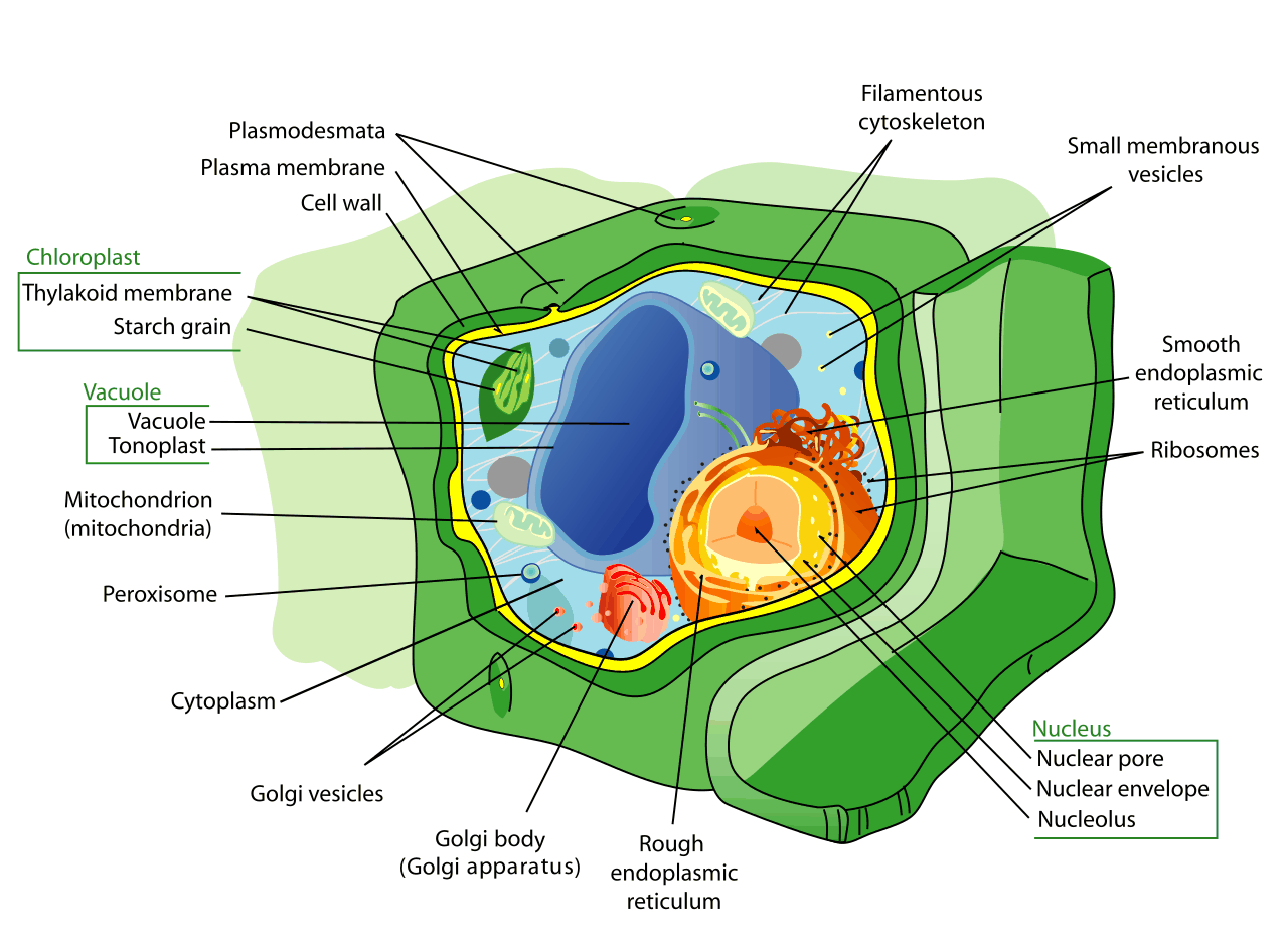
A plant cell has all the organelles of an animal cell except centrioles, plus three more structures:
- Cell wall: made of cellulose; strong and fully permeable; gives shape and support and stops the cell bursting when it takes in water.
- Chloroplasts: oval, with a double membrane, grana and stroma; carry out photosynthesis.
- Large permanent vacuole: filled with cell sap; stores water, sugars and salts and keeps the cell firm (turgid).
| Feature | Plant cell | Animal cell |
|---|---|---|
| Cell wall | Present (cellulose) | Absent |
| Chloroplasts | Present in green parts | Absent |
| Vacuole | One large, permanent, central | Small and temporary, if any |
| Shape | Fixed and regular | Irregular, can change |
| Centrioles | Absent (in flowering plants) | Present |
| Nucleus | Usually pushed to one side | Usually central |
| Stored food | Starch (and oils) | Glycogen (and fats) |
They differ because of how the organisms live. Plants make their own food (so they need chloroplasts), do not move about (so the wall supports them) and take in water by osmosis (so the wall stops bursting). Animals eat ready-made food and move, so a flexible membrane without a wall suits them.
Specialised cells
Specialised cell
A cell whose structure is adapted to carry out one particular function.
| Cell | Where found | Adaptations to its function |
|---|---|---|
| Root hair cell | Surface of young roots | Long hair-like extension gives a large surface area; thin wall; concentrated cell sap draws in water by osmosis |
| Palisade cell | Below the upper epidermis of a leaf | Tall, closely packed; many chloroplasts to absorb light for photosynthesis |
| Guard cell | In pairs around each stoma | Bean-shaped; thick inner wall makes the cells curve and open the stoma when turgid; has chloroplasts |
| Pollen grain | Anthers of flowers | Tough wall protects it; spiky or sticky surface for insects, or light and smooth for wind; carries the male nucleus |
| Muscle cell | Muscles | Long and thin; protein fibres contract; many mitochondria supply energy |
| Nerve cell (neurone) | Brain, spinal cord, nerves | Very long axon carries impulses far; branched dendrites connect with many cells; fatty sheath speeds impulses |
| Red blood cell | Blood | Biconcave disc: large surface area; no nucleus: more room for haemoglobin to carry oxygen; flexible |
| White blood cell | Blood and lymph | Changes shape to engulf germs (phagocytes) or makes antibodies (lymphocytes) |
| Sperm cell | Testes | Tail for swimming; many mitochondria for energy; enzymes in the head to enter the egg |
| Egg cell (ovum) | Ovaries | Large, with stored food for the early embryo |
Levels of organisation
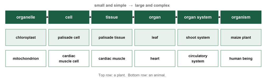
Tissue
A group of similar cells working together to perform one function, for example muscle tissue or xylem.
Organ
A structure made of different tissues working together, for example the heart, the stomach or a leaf.
Organ system
A group of organs working together to carry out a major life process, for example the digestive system.
Order of organisation: organelle → cell → tissue → organ → organ system → organism. Single-celled organisms such as amoeba and yeast carry out every life process inside one cell; in large organisms cells divide the work among themselves.
Key points to remember
- The cell is the basic unit of structure and function of life; cells are measured in micrometres (1 mm = 1,000 micrometres).
- The electron microscope has far higher magnification and resolution than the light microscope but uses only dead specimens.
- Temporary slides are prepared by sectioning, staining and mounting; fixation preserves tissue for permanent slides.
- Cell size = diameter of field of view ÷ number of cells across it; total magnification = eyepiece × objective.
- Plant cells have a cellulose wall, chloroplasts and a large vacuole; animal cells have centrioles and no wall.
- Specialised cells such as root hair, palisade, guard, nerve, muscle and red blood cells are adapted to their functions.
- Organelles → cells → tissues → organs → organ systems → organism.
Practice questions
Distinguish between magnification and resolution. (2 marks)
Show the answer
Magnification is how many times larger the image is than the object; resolution is the ability to show two close points as separate, which decides the detail seen.
Give three differences between the light microscope and the electron microscope. (3 marks)
Show the answer
Any three: light vs electron beam; glass lenses vs electromagnets; about ×1,500 vs ×500,000 or more; low vs high resolution; living or dead vs dead specimens only; colour vs black-and-white image; cheap and portable vs expensive and large.
A learner used a ×10 eyepiece and a ×40 objective. (a) Work out the total magnification. (b) The field of view was 0.4 mm and 5 cells fitted across it. Estimate the length of one cell in micrometres. (3 marks)
Show the answer
- (a) 10 × 40 = ×400.
- (b) 0.4 mm = 400 micrometres; 400 ÷ 5 = 80 micrometres.
Why is iodine solution added when preparing a slide of onion epidermis? (1 mark)
Show the answer
To stain the nucleus (and cytoplasm) so that the parts show clearly.
Why is a cover slip lowered at an angle onto a specimen? (1 mark)
Show the answer
To avoid trapping air bubbles, which would hide parts of the specimen.
State the function of: (a) mitochondrion (b) ribosome (c) Golgi apparatus (d) lysosome. (4 marks)
Show the answer
(a) Aerobic respiration, releasing energy (b) making proteins (c) modifying and packaging proteins for secretion (d) digesting worn-out organelles and engulfed particles.
State three structures found in a plant cell but not in an animal cell, and give the function of each. (6 marks)
Show the answer
- Cell wall: support, shape, prevents bursting.
- Chloroplast: photosynthesis.
- Large permanent vacuole: stores cell sap and keeps the cell turgid.
Explain how a red blood cell is adapted to carrying oxygen. (3 marks)
Show the answer
- Biconcave shape gives a large surface area for oxygen to diffuse in and out.
- No nucleus, so there is more room for haemoglobin.
- Thin and flexible, so it squeezes through capillaries.
Arrange in order from simplest to most complex: organ, cell, organism, tissue, organelle, organ system. (2 marks)
Show the answer
Organelle, cell, tissue, organ, organ system, organism.
This is one topic. Get the whole year. The full Grade 10 Biology notes cover every sub-strand of Terms 1, 2 and 3 like this one, with pictures, key points and practice questions with answers, as a printable PDF for KSh 150. Pay with M-Pesa and download at once.
Get the full Grade 10 Biology notes · All Grade 10 Biology resources · Notes for every grade


